A copper(II)–dipicolylamine–coumarin sensor for maltosyltransferase assay

A Cu(II)−[di(2-methylpyridyl)methylamino]coumarin fluorescence turn-on sensor (Cu-1b) is designed to detect phosphate ion with Kass = 1.4 × 10⁵ M–1 in HEPES buffer. Cu-1b is applied to probe the GlgE catalyzed maltose-transfer reaction of α-maltose-1-phosphate to α-1,4-glucan with concomitant release of phosphate ion in Mycobacterium tuberculosis. Dalton Trans. 2019, 48, 8026–8029.
Quantitative analysis of sugar ingredients in beverages and food crops by an effective method combining naphthimidazole derivatization and 1H-NMR spectrometry

Background: Though carbohydrates are needed for health, excessive uptake of sugar may induce obesity, decayed teeth, and chronic diseases. The Taiwan Food and Drug Administration has recently proposed to regulate
common sugars, a list that includes glucose, galactose, fructose, lactose, maltose, and sucrose in beverages and dietary foods. Accordingly, rapid and effective methods are needed for the quantitative analysis of sugar ingredients
in beverages and foods.
Objective: To establish a convenient method of quantifying sugar ingredients in beverages and foods by using one-dimensional 1H-NMR spectroscopy via a simple treatment with naphthimidazole (NAIM) labeling kit.
Methods: The sample of beverages or the hydrolysate of food crops were treated with an NAIM labeling kit that contains 2,3-naphthalenediamine and iodine in acetic acid solution. After the complete conversion of the reducing sugars to sugar-NAIM derivatives, the mixture solution was subjected to rotary evaporation under reduced pressure, and the residue was dissolved in D2O solution with dimethylsulfoxide (DMSO) as an internal standard for the 1H-NMR spectrometric analysis.
Results: An aldose existing in two anomeric isomers is completely converted to the corresponding NAIM derivative that shows a single characteristic vinyl proton at a distinct position to facilitate the quantitative analysis by NMR spectrometry. Even 10 μmol of common sugar ingredients in 50 μL of beverage or in the hydrolysate prepared from 1 mg of food crop can be quantified.
Conclusions: The results suggest that a simple treatment of beverage with the NAIM labeling kit provides a convenient method for the quantification of sugar ingredients by 1H-NMR spectrometry. This method combining NAIM derivatization and 1H-NMR analysis is also useful for the profiling and fingerprinting of food crops. Functional Foods in Health and Disease, 2017, 7, 494-510
A cell-permeable and triazole-forming fluorescent probe for glycoconjugate imaging in live cells

A new fluorescence-forming probe, coumOCT, designed by fusing cyclooctyne with a coumarin fluorophore was successfully used for the imaging of azido-glycoconjugates in living HeLa cells. This probe is cell-permeable and generates fluorescence after triazole formation, thus minimizing the background signal and enabling the real-time intracellular imaging of glycoconjugate trafficking. Chem. Commun., 2017,53, 1490–1493
Fluorescent Sensing of Guanine and Guanosine Monophosphate with Conjugated Receptors Incorporating Aniline and Naphthyridine Moieties

Ethyne-linked naphthyridine–aniline
conjugated molecules are selective sensors of decylguanine in dichloromethane and guanosine monophosphate in water (Kass = 16 000 M–1). The 2-acetamido-1,8-naphthyridine moiety binds with guanine in a DAA–ADD
triply hydrogen-bonded motif. The aniline moiety enhances an electron-donating effect, and the substituent is tuned to attain extra hydrogen bonds, π–π stacking, and electrostatic interactions. The proposed binding modes are
supported by a Job plot, ESI-MS, 1H NMR, UV–vis, and fluorescence spectral analyses.
Org. Lett. 2016, 18, 1724−1727
Tracking and Finding Slow-Proliferating/Quiescent Cancer Stem Cells with Fluorescent Nanodiamonds
Quiescent cancer stem cells (CSCs) have long been considered to be a source of tumor initiation. However, identification and isolation of these cells have been hampered by the fact that commonly used fluorescent markers are not sufficiently stable, both chemically and photophysically, to allow tracking over an extended period of time. Here, it is shown that fluorescent nanodiamonds (FNDs) are well suited for this application. Genotoxicity tests of FNDs with comet and micronucleus assays for human fibroblasts and breast cancer cells indicate that the nanoparticles neither cause DNA damage nor impair cell growth. Using AS-B145-1R breast cancer cells as the model cell line for CSC, it is found that the FND labeling outperforms 5-ethynyl-2′-deoxyuridine (EdU) and carboxyfluorescein diacetate succinimidyl ester (CFSE) in regards to its long-term tracking capability (>20 d). Moreover, through a quantification of their stem cell activity by measuring mammosphere-forming efficiencies (MFEs) and self-renewal rates, the FND-positive cells are identified to have an MFE twice as high as that of the FND-negative cells isolated from the same dissociated mammospheres. Thus, the nanoparticle-based labeling technique provides an effective new tool for tracking and finding slow-proliferating/quiescent CSCs in cancer research. Small 2015, 11, 4394–4402.
New Continuous Fluorometric Assay for Bacterial Transglycosylase Using Förster Resonance Eenergy Ttransfer

The emergence of antibiotic resistance has prompted scientists to search for new antibiotics. Transglycosylase (TGase) is an attractive target for new antibiotic discovery due to its location on the outer membrane of bacteria and its essential role in peptidoglycan synthesis. Herein, we report a new continuous fluorescent assay based on Förster resonance energy transfer, using lipid II analogues with a dimethylamino-azobenzenesulfonyl quencher in the lipid chain and a coumarin fluorophore in the peptide chain. During the process of transglycosylation, the quencher-appended polyprenol is released and the fluorescence of coumarin can be detected. This new continuous fluorescent assay not only provides an efficient and convenient way to study TGase activities, but also enables the high-throughput screening of potential TGase inhibitors for antibiotic discovery. J. Am. Chem. Soc. 2013, 135, 17078–17089.
Chemical Probes for Drug-Resistance Assessment by Binding Competition (RABC): Oseltamivir Susceptibility Evaluation
For treatment of influenza infections, neuraminidase (NA) inhibitors oseltamivir and zanamivir are the most useful therapies. NAs can evolve to resist OS with mutations such as H274Y, but zanamivir is effective to H274Y mutant and most influenza viruses. The worldwide surge of drug-resistant influenza viruses has prompted the need for development of quick tests for OS susceptibility. We conceive a new method, namely Resistance Assessment by Binding Competition (RABC) assay, using labeled zanamivir as the chemical probe to identify the influenza subtypes that are resistant to OS but still sensitive to zanamivir. This detection platform can be further developed to a quick membrane-based test to enable visual detection, or combined with sugar array analysis for simultaneous determination of drug resistance and virus serotypes. Angew. Chem. Int. Ed. 2013, 52, 366–370.
Nanowire Transistor-based Ultrasensitive Virus Detection with Reversible Surface Functionalization
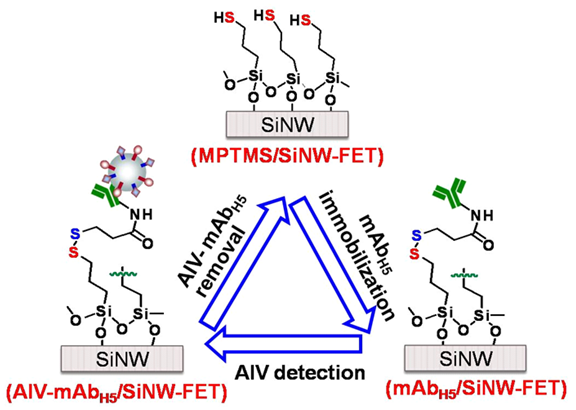
The surface of a 3-mercaptopropyltrimethoxysilane (MPTMS)/SiNW-FET was modified with mAbH5 to become mAbH5/SiNW-FET through a disulfide linker. After the AIV detection, the AIV-mAbH5 complex on the SiNW-FET was removed by dithiothreitol (DTT) washing to return the device to the MPTMS/SiNW-FET. Chem. Asian J. 2012, 7, 2073–2079.
Rapid and Specific Influenza Virus Detection by Functionalized Magnetic Nanoparticles and Mass Spectrometry

The timely and accurate diagnosis of specific influenza virus strains is crucial to effective prophylaxis, vaccine preparation and early antiviral therapy. In conjugation with the immunoassay utilizing monoclonal antibody, mass spectrometry is an alternative to identify proteins derived from a target influenza virus. Taking advantage of the large surface area-to-volume ratio, antibody-conjugated magnetic nanoparticles can act as an effective probe to extract influenza virus for sodium dodecylsulfate polyacrylamide gel electrophoresis (SDS-PAGE) and on-bead mass spectrometric analysis. J. Nanobiotech. 2011, 9, 52.
Synthesis and Evaluation of a New Fluorescent Transglycosylase Substrate: Lipid II-based Molecule Possessing a Dansyl-C20 Polyprenyl Moiety

A novel fluorescent lipid II-based substrate for transglycosylases (TGases) is designed, via an imaginary transform A, to incorporate a dansyl group to mimic the diprenyl moiety in the lipid chain. This fluorescent substrate is readily utilized in the functional assay of TGases without an additional treatment with another enzyme of N-acetylmurmidase. Org. Lett. 2010, 12, 1608–1611.
A Fluorescence Sensor for Detection of Geranyl Pyrophosphate by Chemo-Ensemble Method

A chemo-ensemble system using FRET as the sensing mechanism to detect isoprenoid pyrophosphates. The hexaamide receptor molecule has a well-defined structure with pseudo-tetrahedral cleft to accommodate the pyrophosphate, and incorporates two long hydrocarbon chains to enhance the interactions with the aliphatic moiety of GPP. The coumarin phosphate is selected as an appropriate fluorescence indicator because it binds modestly with the receptor to render a FRET from the pyrene rings. GPP selectively replaces coumarin phosphate, but other anions including CH3CO2–, C10H21CO2–, C11H23CO2–, F–, Cl–, Br–, SCN–, NO3–, and ClO4– cannot. This chemo-ensemble system provides an efficient, selective and sensitive method for detection of GPP. J. Org. Chem. 2009, 74, 895–898.
Phospholipid-Induced Aggregation and Anthracene Excimer Formation

The zinc complex of anthryl bis(dipicolylamine) ( 1 ) aggregates upon binding with long-chain aliphatic phosphates and displays anthracene excimer fluorescence, which provides a new strategy toward detection of the biologically important lysophosphatidic acid in aqueous solution. Org. Lett. 2008, 10, 4401-4404.
Production of Antibodies for Selective Detection of Malachite Green and the Related Triphenylmethane Dyes in Fish and Fishpond Water

This study provides a practical method for production of the antibodies against malachite green (MG) and its primary metabolite leucomalachite green (LMG). Two ELISA kits are constructed with the MG and LMG antibodies for detection of the residual MG and LMG in fish muscle and fishpond water. The detection limit is established at the level of 0.05 μg/L for both MG and LMG. Our ELISA kits show the advantages of good specificity, high sensitivity, and convenience in rapid screening of MG and LMG residues. The sample of fishpond water, without extraction or prior preparation, is directly assayed by the ELISA kit. More then 80 fish samples can be simultaneously tested in a kit. The toxic crystal violet and its metabolite leucocrystal violet of illegal use in aquaculture are detected by our prepared MG and LMG antibodies, whereas the antibodies do not cross-react with common antibiotics, sulfonamides, and benzene derivatives. J. Agric. Food Chem.2007, 55, 8851.
Fluorescent and Circular Dichroic Detection of Monosaccharides by Molecular Sensors: Bis[(Pyrrolyl)ethynyl]naphthyridine and Bis[(Indolyl)ethynyl]naphthyridine
ethynyl]naphthyridine and Bis[(Indolyl)ethynyl]naphthyridine.jpg)
The push - pull conjugated molecules BPN and BIN adopting daad relays of proton donors(d) and acceptors(a) form multiple hydrogen-bonding complexes with various monosaccharides that possess complementary adda sequences. On the other hand, the CD-silent BIN molecule binds with monosaccharides to form the CD-active multiple hydrogen- bonding complexes, which exhibit the remarkable chirality dependent helicities consistent with the prediction by the ab initio approaches. J. AM. CHEM. SOC. 2004, 126, 3559–3566.