- The full article entitled “Wood cellulose microfibrils have a 24-chain core–shell nanostructure in seed plants” can now be found at the Nature Plants website at https://www.nature.com/articles/s41477-023-01430-z
- Authors: Hwan-Chin Tai*, Chih-Hui Chang, Wenjie Cai, Jer-Horng Lin, Shing-Jong Huang, Qian-Yan Lin, Eric Chung-Yueh Yuan, Shu-Li Li, Ying-Chung Jimmy Lin, Jerry Chun Chung Chan*, and Cheng-Si Tsao*
Wood is a crucial foundational material and energy source in human history, representing a sustainable and renewable resource for the future. Cellulose in the wood cell walls makes up half of its weight, making it one of the most abundant organic substances on Earth, accounting for a quarter of the global biomass. Despite a century-long history of studying the structure of wood cellulose, its nanoscale structure remains unresolved. Specifically, the number of cellulose chains within each cellulose microfibril bundle is a contentious topic. While electron and atomic force microscopes have the capability to analyze single molecular chains, they cannot differentiate cellulose from the surrounding encapsulating hemicellulose, which prevents a direct calculation of microfibril chain counts. As far back as 1913, Professor Shogi Nishikawa used small-angle X-ray scattering (SAXS) to observe that the molecular arrangement of wood cellulose exhibited directionality. However, over the past century, a comprehensive theoretical model for quantitative analysis has been lacking. Under the leadership of Professor Hwan-Chin Tai, a former PI at the Department of Chemistry, NTU, this study significantly enhanced the measurement signals for wood using the BioSAXS beamline at Taiwan Photon Source, a third-generation synchrotron facility. Professor Cheng-Si Tsao, a joint-appointed professor in the Department of Materials Science and Engineering at National Taiwan University, developed a novel model that incorporated considerations of length effects into the SAXS analysis mode. If the proportion of cellulose molecules in the wood's core and shell regions is known, the calculation of the cellulose chain count in the core area can be achieved through fitting. Although, in principle, one could measure the proportion of cellulose in different regions using C-13 solid-state nuclear magnetic resonance spectroscopy, the significant overlap between hemicellulose and cellulose carbon-13 signals has led to substantial measurement inaccuracies in this critical molecular proportion. To address this challenge, Professor Jerry Chan's team from the Department of Chemistry developed the GIFTED (Global Iterative Fitting of T1ρ-Edited Decay) pulse sequence. This technique exploits the differences in the movement of cellulose and hemicellulose molecules, resulting in different decay rates under spin-locking conditions, greatly enhancing the accuracy of the cellulose proportion between the core and shell regions. By combining these two innovative techniques, this study successfully estimated the cellulose chain count in wood microfibrils, unexpectedly arriving at a conclusion of 24 chains, overturning the most commonly cited 36-chain model found in textbooks for decades. This achievement solves the century-long challenge of understanding cellulose structure in wood cell walls, bearing significant implications for the future sustainable utilization of wood through genetic improvement and cellulose resources. Additionally, the GIFTED technique can be widely applied to study amorphous samples in natural abundance, such as polymers, partially alleviating the issue of insufficient C-13 spectral resolution.
Chih-Hui Chang, a graduate student from the Department of Chemistry at National Taiwan University, is the first non-corresponding author. The GIFTED technique was jointly developed by Qian-Yan Lin, Eric Chung-Yueh Yuan, and Shu-Li Li.
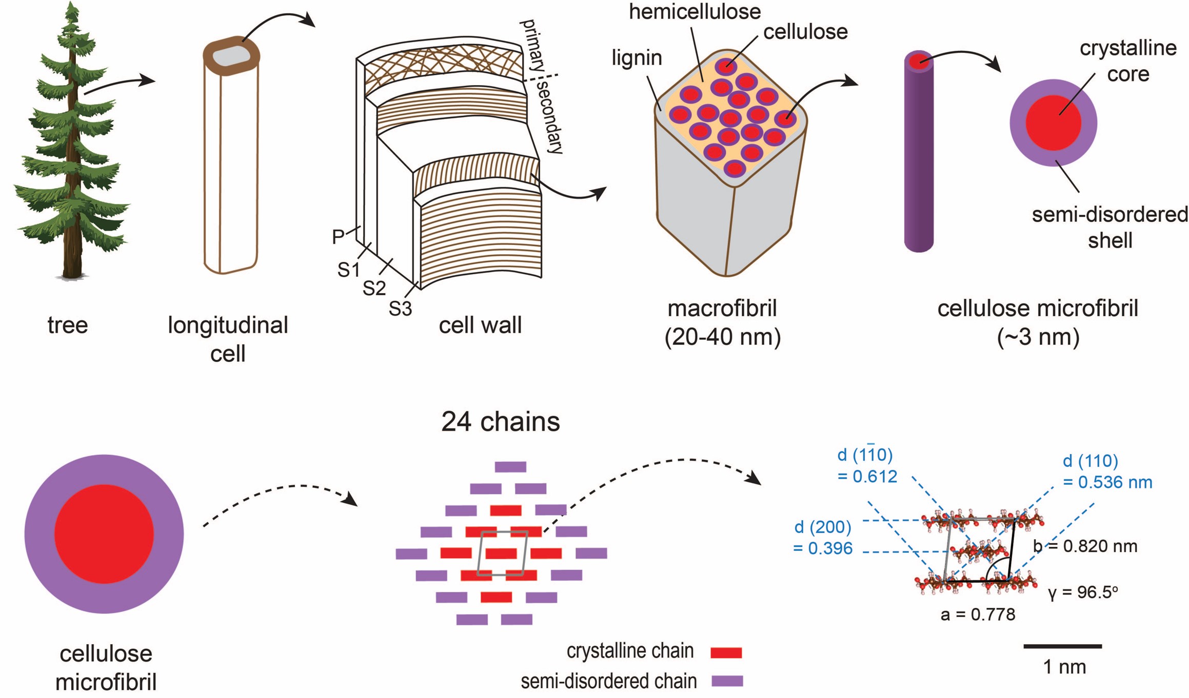
Figure 1: Schematics of Wood Structure. Wood cellulose is primarily found in the S2 layer of the longitudinal secondary cell wall, forming nanoscale microfibrils. These research results confirm for the first time that each microfibril bundle contains 24 cellulose chains, exhibiting a shell-core structure.